43 light microscope with labels
Microscope labels Flashcards | Quizlet Microscope label Learn with flashcards, games, and more — for free. Microscope Labeling - The Biology Corner 1) Start with scanning (the shortest objective) and only use the COARSE knob . Once it is focused… 2) Switch to low power (medium) and only use the COARSE knob . You may need to recenter your slide. Once it is focused.. 3) Switch to high power (long objective).
Compound Microscope Parts - Labeled Diagram and their Functions The eyepiece (or ocular lens) is the lens part at the top of a microscope that the viewer looks through. The standard eyepiece has a magnification of 10x. You may exchange with an optional eyepiece ranging from 5x - 30x. [In this figure] The structure inside an eyepiece. The current design of the eyepiece is no longer a single convex lens.

Light microscope with labels
Light Microscope: Functions, Parts and How to Use It The function of the light microscope is based on its ability to focus a beam of light through a very small and transparent specimen, to produce an image. The image is then passed through one or two lenses for magnification to view. The transparency of the specimen allows for easy and fast light penetration. Specimens can vary from bacteria to ... rsscience.com › stereo-microscopeParts of Stereo Microscope (Dissecting microscope) - Rs' Science Labeled part diagram of a stereo microscope Major structural parts of a stereo microscope. There are three major structural parts of a stereo microscope. The viewing Head includes the upper part of the microscope, which houses the most critical optical components, including the eyepiece, objective lens, and light source of the microscope. Light Microscope- Definition, Principle, Types, Parts, Labeled Diagram ... A light microscope is a biology laboratory instrument or tool, that uses visible light to detect and magnify very small objects and enlarge them. They use lenses to focus light on the specimen, magnifying it thus producing an image. The specimen is normally placed close to the microscopic lens.
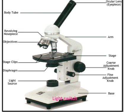
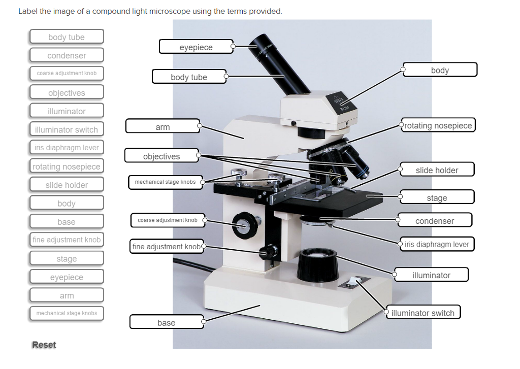
Light microscope with labels. PDF Parts of the Light Microscope - Science Spot Supports the MICROSCOPE D. STAGE CLIPS HOLD the slide in place C. OBJECTIVE LENSES Magnification ranges from 10 X to 40 X F. LIGHT SOURCE Projects light UPWARDS through the diaphragm, the SPECIMEN, and the LENSES H. DIAPHRAGM Regulates the amount of LIGHT on the specimen E. STAGE Supports the SLIDE being viewed K. ARM Used to SUPPORT the Light Microscope - an overview | ScienceDirect Topics The light microscope is an instrument for visualizing fine detail of an object. It does this by creating a magnified image through the use of a series of glass lenses, which first focus a beam of light onto or through an object, and convex objective lenses to enlarge the image formed. › products › microscopeMicroscope Objective Lens | Products | Leica Microsystems The objective lens is a critical part of the microscope optics. The microscope objective is positioned near the sample, specimen, or object being observed. It has a very important role in imaging, as it forms the first magnified image of the sample. The numerical aperture (NA) of the objective indicates its ability to gather light and largely determines the microscope’s resolution, the ... Compound Light Microscopes | Products | Leica Microsystems Light microscope solutions help suppliers and device manufacturers achieve fast and precise inspection and analysis for semiconductor wafer processing. Conformity to the defined specifications during semiconductor device manufacturing is critical for reliability. ... (CRS) is a powerful approach for label-free, chemically specific imaging. It ...
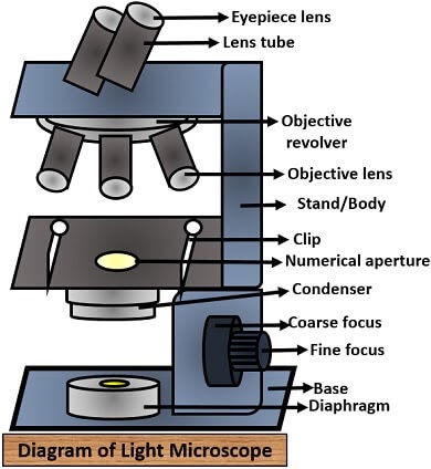
Parts of a microscope with functions and labeled diagram Microscopic illuminator - This is the microscopes light source, located at the base. It is used instead of a mirror. It captures light from an external source of a low voltage of about 100v. Condenser - These are lenses that are used to collect and focus light from the illuminator into the specimen. Parts of the Microscope with Labeling (also Free Printouts) Parts of the Microscope with Labeling (also Free Printouts) A microscope is one of the invaluable tools in the laboratory setting. It is used to observe things that cannot be seen by the naked eye. Table of Contents 1. Eyepiece 2. Body tube/Head 3. Turret/Nose piece 4. Objective lenses 5. Knobs (fine and coarse) 6. Stage and stage clips 7. Aperture Labelled Diagram Of A Light Microscope - GlobalSpec Products/Services for Labelled Diagram Of A Light Microscope Microscopes - (706 companies) ...and transmission electron microscopes. Acoustic and ultrasonic microscopes use sound waves to create images of the sample. Compound microscopes use a single light path. These types of microscopes can have a single eyepiece (monocular) or a dual eyepiece... Microscope Drawing And Label - Painting Valley Label The Microscope... 270x350 14 0 Compound Light Micro... 630x380 8 1 Labeling The Parts O... 525x450 7 0 Compound Microscope ... 413x424 6 0 Microscope - Microsc... 236x262 4 1 Label Microscope Dia... 459x457 4 0 Microscope Parts Dia... 576x400 3 0 Section Cells View A... 512x346 2 1 Exe - Microscope Dra... 933x1163 2 0 Drawing - Microscope...
Microscope Parts and Functions Microscope Parts and Functions With Labeled Diagram and Functions How does a Compound Microscope Work?. Before exploring microscope parts and functions, you should probably understand that the compound light microscope is more complicated than just a microscope with more than one lens.. First, the purpose of a microscope is to magnify a small object or to magnify the fine details of a larger ... Microscope, Microscope Parts, Labeled Diagram, and Functions Majority of high quality microscopes used in laboratory include an Abbe condenser with an iris diaphragm. When iris diaphragm is combined with Abbe condenser, it control both the quantity of light applied as well as focus on the specimen. Aperture: It is the hole in the stage through which the base (transmitted) light reaches the stage. Compound Microscope Parts, Functions, and Labeled Diagram Compound Microscope Definitions for Labels. Eyepiece (ocular lens) with or without Pointer: The part that is looked through at the top of the compound microscope. Eyepieces typically have a magnification between 5x & 30x. Monocular or Binocular Head: Structural support that holds & connects the eyepieces to the objective lenses. Microscope Labeling - The Biology Corner Students label the parts of the microscope in this photo of a basic laboratory light microscope. Can be used for practice or as a quiz. Name_____ Microscope Labeling . Microscope Use: 15. When focusing a specimen, you should always start with the _____ objective.
BIO 156, Fall 2015: Week 8. Lab 7. Immune System
Animal Cell Under Light Microscope Labelled : Draw and label the ... Most cells are visible under a light microscope, but mitochondria and bacteria are barely visible. Record the microscope images using labelled diagrams or produce digital images. We say cells are microscopic because they can only be seen under a microscope. Note the whiplike flagellum that gives the cell a threadlike appearance.
Using a Light Microscope - AyushiSinhaMicroscopy
› microscopy › intSmart Microscope for Lab Routine and Research - ZEISS Acquiring fluorescent images has never been so easy. Combine Axioscope 5 with the LED light source Colibri 3 and the sensitive, standalone microscope camera Axiocam 202 mono to have the perfect setup for easy multichannel fluorescence documentation. Switch effortlessly between the channels for UV, blue, green and red excitation.

Unlabeled Compound Light Microscope Diagram - Micropedia
Label the Light Microscope - Labelled diagram - Wordwall Eyepiece, Light Source, Base, Stage, Stage Clips, Fine Focus, Coarse Focus, Arm, Objective Lens. ... Label the Light Microscope. Share Share by Nquinn805. Like. Edit Content. Embed. More. Leaderboard. Show more Show less . This leaderboard is currently private. Click Share to make it public. This leaderboard has been disabled by the resource ...
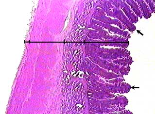
Duodenum
Light Microscope : Main Parts of Light Microscope | Biology It is a simple ordinary microscope which is most popularly used in biological studies. The first light microscope was made by Janssen and Hans in 1590. The light microscope can magnify objects upto 2000 times. Its limits of resolution are about 0.2 µ, about one-half the wavelength of the light used.
A Light Microscope - Micropedia
Animal Cell Diagram Under Light Microscope Labeled Tuesday, April 20th 2021. | Diagram. Animal Cell Diagram Under Light Microscope. To make observations and draw scale. This shows a generalized animal cell under a light microscope. We all keep in mind that the human physique is amazingly elaborate and one way I discovered to comprehend it is by way of the style of human anatomy diagrams.
Search in gallery
Light Microscope- Definition, Principle, Types, Parts, Labeled Diagram ... A light microscope is a biology laboratory instrument or tool, that uses visible light to detect and magnify very small objects and enlarge them. They use lenses to focus light on the specimen, magnifying it thus producing an image. The specimen is normally placed close to the microscopic lens.

Compound Light Microscope Parts And Functions Worksheet | Decoratingspecial.com
rsscience.com › stereo-microscopeParts of Stereo Microscope (Dissecting microscope) - Rs' Science Labeled part diagram of a stereo microscope Major structural parts of a stereo microscope. There are three major structural parts of a stereo microscope. The viewing Head includes the upper part of the microscope, which houses the most critical optical components, including the eyepiece, objective lens, and light source of the microscope.

Bone Lab
Light Microscope: Functions, Parts and How to Use It The function of the light microscope is based on its ability to focus a beam of light through a very small and transparent specimen, to produce an image. The image is then passed through one or two lenses for magnification to view. The transparency of the specimen allows for easy and fast light penetration. Specimens can vary from bacteria to ...

Microscope With Labels vector, free vector graphics - Vector.me
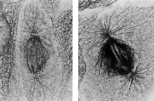
Nanogold-Antibody Conjugates

Microscope Filter Cubes
302 Found
Using the Microscope
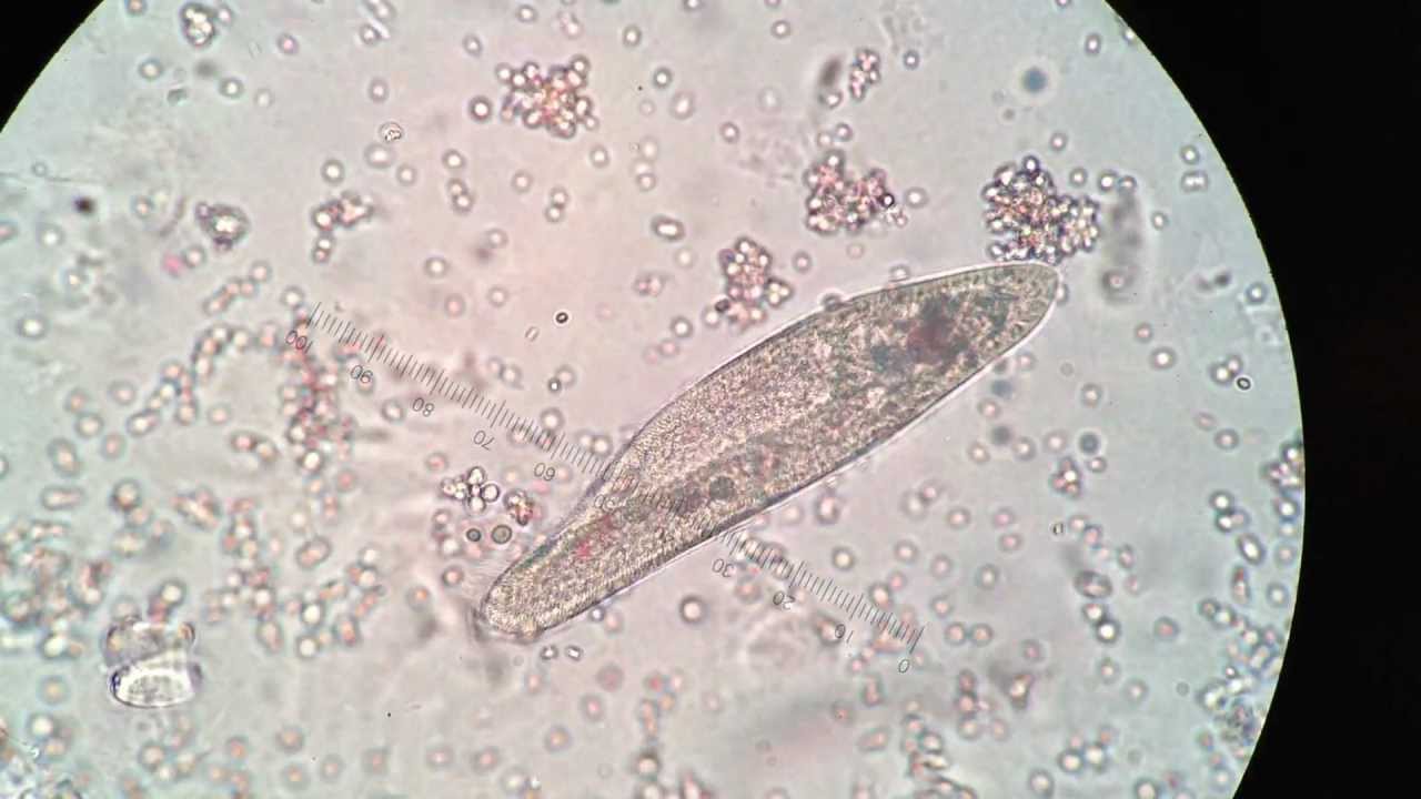
Paramecium under 400X magnification - YouTube

Types Of Light Microscopes
Solved: Label The Image Of A Compound Light Microscope Usi... | Chegg.com

Euglena Acus 2 - BF microscope 1250x - YouTube
Post a Comment for "43 light microscope with labels"