41 spinal cord model with labels
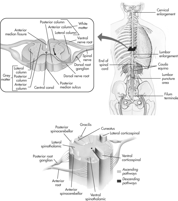
Spinal cord: Anatomy, structure, tracts and function | Kenhub The spinal cord is made of gray and white matter just like other parts of the CNS. It shows four surfaces: anterior, posterior, and two lateral. They feature fissures (anterior) and sulci (anterolateral, posterolateral, and posterior). The gray matter is the butterfly-shaped central part of the spinal cord and is comprised of neuronal cell bodies. Anatomy of the spinal cord - e-Anatomy - IMAIOS This atlas of human anatomy describes the spinal cord through 18 anatomical diagrams with 270 anatomical structures labeled. It was designed particularly for physiotherapists, osteopaths, rheumatologists, neurosurgeons, orthopedic surgeons and general practitioners, especially for the study and understanding of medullary diseases.
Spinal cord transverse section coverings label 3D model - CGTrader Spinal cord transverse section coverings label 3D model, available formats , ready for 3D animation and other 3D projects | CGTrader.com ... A blend model of spinal cord along with it covering layers and nerve roots. The ascending and descending tracts of spinal cord transverse section are labelled in detail. The material is image textures with ...

Spinal cord model with labels
Learn the spinal cord with diagrams and quizzes | Kenhub The spinal cord, along with the brain, makes up the central nervous system (CNS). It is a long tubular structure comprised of nervous tissue, extending from the cervical to the lumbar region of the vertebral column. Just like other parts of the CNS, the spinal cord is comprised of white and gray matter. Spinal cord gray matter is the central ... Labeled Brain Model Diagram | Science Trends The medial region of the posterior and anterior lobes function to control fine body movements, taking in input from the spinal cord as well as the auditory and visual systems of the brain. The lateral region of the cerebellum is the largest part of the cerebellum in humans. This region gets inputs from the cerebral cortex. Fibroblast Growth Factor Receptor Signaling in ... May 09, 2012 · As seen in the spinal cord, myelin thickness was also reduced in the optic nerves of the mutants compared with controls. Quantification of myelin thickness by g-ratio analysis (expressed as scatter plots) confirmed the reduction of myelin thickness in the mutants (average g-ratios: controls, 0.78 ± 0.05; mutants, 0.85 ± 0.06; p = 7.4 × 10 ...
Spinal cord model with labels. PDF Spinal Cord Classroom Teacher - Duquesne University after reviewing the different parts of the spinal cord, students will draw their own representations of the spinal cord. remind students to include labels on their drawings showing some of the different parts of the spinal cord they learned about! 3. 4. Use spools and thread to make a spinal cord. B o n e M o d u l e t e a C h e r W p a g e S Spinal Cord Model - YouTube Going over things we need to know for our practical... spinal cord lab model Diagram | Quizlet spinal cord lab model Diagram | Quizlet spinal cord lab model STUDY Learn Write Test PLAY Match + − Created by jasmein_rice Terms in this set (8) anterior horn ... ventral root ... lateral horn ... dorsal root ganglion ... spinal nerve ... posterior horn ... central canal ... dorsal root ... Sets found in the same folder sensory system 79 terms SPINAL CORD MODEL Flashcards | Quizlet Objectives for Spinal Cord (fifth cervic…. 210: Chapter 11 Blended Skills and Critical Thinki…. 5th - Social Studies Review - ch7 notes and questi….
spinal cord anatomy, labeling spinal model Quiz This is an online quiz called spinal cord anatomy, labeling spinal model There is a printable worksheet available for download here so you can take the quiz with pen and paper. Your Skills & Rank Total Points 0 Get started! Today's Rank -- 0 Today 's Points One of us! Game Points 16 You need to get 100% to score the 16 points available Actions The FASEB Journal - Wiley Online Library The FASEB Journal publishes high quality and impactful multidisciplinary research covering biology and biomedical sciences at every level of organization: atomic, molecular, cell, tissue, organ, organismic, and population. PDF Anatomy and Physiology of the Spinal Cord The spinal cord is a bundle of spinal nerves wrapped together. The spinal nerves enter and exit the spinal cord through small spaces between the vertebrae. The blood vessels which carry oxygen to the spinal cord also use these spaces. You have 8 pairs of cervical nerves, 12 thoracic, 5 lumbar and 6 sacral. Spinal Cord Injury | Types of Spinal Cord Injuries ... 88 percent of spinal cord injury survivors who were single at the time of the accident are single five years later, compared to 65 percent in the general population. Two-thirds of sports-related spinal cord injuries are from diving, making it the most dangerous sport for the brain and spinal cord.
The spinal cord | Human Anatomy and Physiology Lab (BSB 141) | | Course ... The spinal cord Information The spinal cord in cross-section has a central region of darker gray matter and the rest is lighter white matter. The gray matter is made up of neuroglia cells and neuron cell bodies. The white matter is made up of neuron axons, mostly but not all myelinated. Nervous System Models | Spinal Cord with Nerves Models Deluxe Spinal Cord Model (0165-00) Item # DGA65. $369.00 $339.00. Add to cart. Kyoto Kagaku Full-Figure Nervous System Model. Item # KK-A25. Add to cart. Kyoto Kagaku Nerves and Vessels of Arm Model. Item # KK-A144. Add to cart. Physiology of Nerves Series, 5 magnetics - illustrated metal board ... The Brain and Nervous System | Noba It begins as a simple bundle of tissue that forms into a tube and extends along the head-to-tail plane becoming the spinal cord and brain. 25 days into its development, the embryo has a distinct spinal cord, as well as hindbrain, midbrain and forebrain (Stiles & Jernigan, 2010). What, exactly, is this nervous system that is developing and what ... Nervous System Models - Labeled Brain and Spinal Cord - Pinterest Brain Model Labeled C Carmel Moore Nursing and Medical Muscle Anatomy Spinal Cord Protection Just like the brain, the spinal cord is covered in both meninges and bone. The bone is what you'd typically think of as being the "spine". The meninges have the same names and... L Leslie Smart Physical Therapy Anatomy Humor Anatomy Coloring Book
Spinal cord transverse section coverings label 3D Model Model's Description: Spinal cord transverse section coverings label 3d model contains 2,035,824 polygons and 835,146 vertices. Please wait for full texture to load (HD). A blend model of spinal cord along with it covering layers and nerve roots. The ascending and descending tracts of spinal cord transverse section are labelled in detail.
PDF Anatomy & Physiology - TMCC Somso Model KS 4 Block model showing the skin with hair in different planes of section. I. Epidermis II. Corium (Dermis) III. Subcutis (Hypodermis) 1. External Horny Layer (Stratum corneum) 1a. Clear Layer (Stratum lucidum) -(KS 3 only) 2. Internal Hornless Germinative Zone (Stratum germinativum) 2a. Granular Layer (Stratum granulosum) 2b.
9,901 Spinal Cord Stock Photos and Images - 123RF Model of a human spine, spinal columns X-ray C-SPINES : AP, LATERAL showing S/p internal fixation C4, C5 & C6 with plate & screws. There is hypersignal intensity lesion in the spinal cord at C4 to C6 levels, probably myelopathy from compression as described above. Spinal segment with a disk
Spinal Cord Models - San Diego Mesa College Spinal Cord Models. Click on a photo for a larger view of the model. Click on Label for the labeled model. Back to Nervous System. Spinal Cord (transverse section) Spinal Cord (close up) Spinal Cord (longitudinal view) Label: Label: Label: Spinal Cord (superior ls) Spinal Cord (inferior ls)
Spinal Cord in the Spinal Canal (BS 31) · Anatomy models | SOMSO® Spinal Cord in the Spinal Canal Seen from the ventral side, natural size, in SOMSO-PLAST®. The model shows the brain stem and the spinal cord, as well as the nerve branches, up to the coccygeal plexus. On the left side, the sympathetic trunk with its connections to the central nervous system is shown. In one piece. Mounted on a green board.
Spinal Cord Diagram with Detailed Illustrations and Clear Labels The spinal cord is one of the most important structures in the human body. In fact, it is the most important structure for any vertebrates. Anatomically, the spinal cord is made up is made up of nervous tissue and is integrated into the spinal column of the backbone. Main Article: Spinal Cord – Anatomy, Structure, Function, and Spinal Cord Nerves
Axis Scientific Spine Model, 34" Life Size Spinal Cord Model with ... This item: Axis Scientific Spine Model, 34" Life Size Spinal Cord Model with Vertebrae, Nerves, Arteries, Lumbar Column, and Male Pelvis, Includes Stand, Detailed Product Manual and Worry Free 3 Year Warranty $76.99 Get it as soon as Friday, Jun 24 FREE Shipping on orders over $25 shipped by Amazon
Solved Fifth vertebra with spinal cord model Labels for the | Chegg.com Science; Biology; Biology questions and answers; Fifth vertebra with spinal cord model Labels for the Fifth vertebra with spinal cord model body of vertebra ventral funiculus ventral root lateral funiculus dorsal root dorsal funiculus dorsal root ganglion gray commissure anterior median fissure central canal: opening posterior median sulcus ventral horn epidural space lateral hom dorsal hom
How to tighten loose skin: 6 tips - Medical News Today Oct 03, 2019 · The effect of oral collagen peptide supplementation on skin moisture and the dermal collagen network: Evidence from an ex vivo model and randomized, placebo‐controlled clinical trials. https ...
Mueller Sports Medicine Adjustable Back Brace, Back Support ... About this item . INTENDED USE: Helps relieve lower back pain while providing support to strains, sprained, and aching backs. FIT: One size. Tapered design provides a comfortable fit for men and women.
Spinal Cord Model - YouTube Dixon discusses Enlargements, cord, pia, dura, mater, denticulate ligaments, 8 spinal nerves, cauda equina, sympathetic chain ganglion , paravetrebral gangli...
Spinal Cord Quiz: Cross-Sectional Anatomy - GetBodySmart Spinal Cord - Cross-Sectional Anatomy. Start Quiz. Want to learn faster? Look no further than these interactive, exam-style anatomy quizzes. Learn anatomy faster and remember everything you learn. Start Now. Related Articles. Parts of the Brain Quiz. Test your knowledge with the parts of the brain and their functions in a fun and interactive ...
Spinal Cord | Trunk Wall (Part 1) | Vertebral Column | Bones of the ... The meninges are the layers of tissue that surround the brain and spinal cord. You can see the meninges on the spinal cords. The dura mater is the tough outer layer, and the arachnoid mater is the thin, transparent layer, deep to the dura mater. The final layer, directly touching the spinal cord, is the pia mater.
Anatomy of the Spinal Cord (Section 2, Chapter 3) Neuroscience Online ... The spinal cord extends from the foramen magnum where it is continuous with the medulla to the level of the first or second lumbar vertebrae. It is a vital link between the brain and the body, and from the body to the brain. The spinal cord is 40 to 50 cm long and 1 cm to 1.5 cm in diameter. Two consecutive rows of nerve roots emerge on each of ...
Labeled Human Torso Model Diagram / torso model anatomy labeled 6678046 orig - Top Label Maker ...
Spinal Cord Labeling Quiz - PurposeGames.com This is an online quiz called Spinal Cord Labeling. There is a printable worksheet available for download here so you can take the quiz with pen and paper. Your Skills & Rank. Total Points. 0. Get started! Today's Rank--0. Today 's Points. One of us! Game Points. 12. You need to get 100% to score the 12 points available.
Spinal Cord - Anatomy, Structure, Function, & Diagram In adults, the spinal cord is usually 40cm long and 2cm wide. It forms a vital link between the brain and the body. The spinal cord is divided into five different parts. Sacral cord Lumbar cord Thoracic cord Cervical cord Coccygeal Several spinal nerves emerge out of each segment of the spinal cord.


Post a Comment for "41 spinal cord model with labels"